

TISSUE MICRO ARRAYS
EXPLORE OUR STATE OF THE ART
AND SPECIALTY ANTIBODIES
Discover a vast collection of normal and diseased tissues, meticulously arrayed and mounted on slides.
Perfect for efficient target characterization in high throughput sample analysis.


Our tissue microarray slides provide the capability to perform rapid analysis of comprehensive panels of normal and disease specimens. Also, for follow-on research, you have the option to access the source tissues for further studies, such as whole sections. Our tissue microarrays are useful in the validation of new gene, protein, and antibody targets. With the matched follow-up and outcome data, a researcher can organize and analyze results in a statistically significant manner.

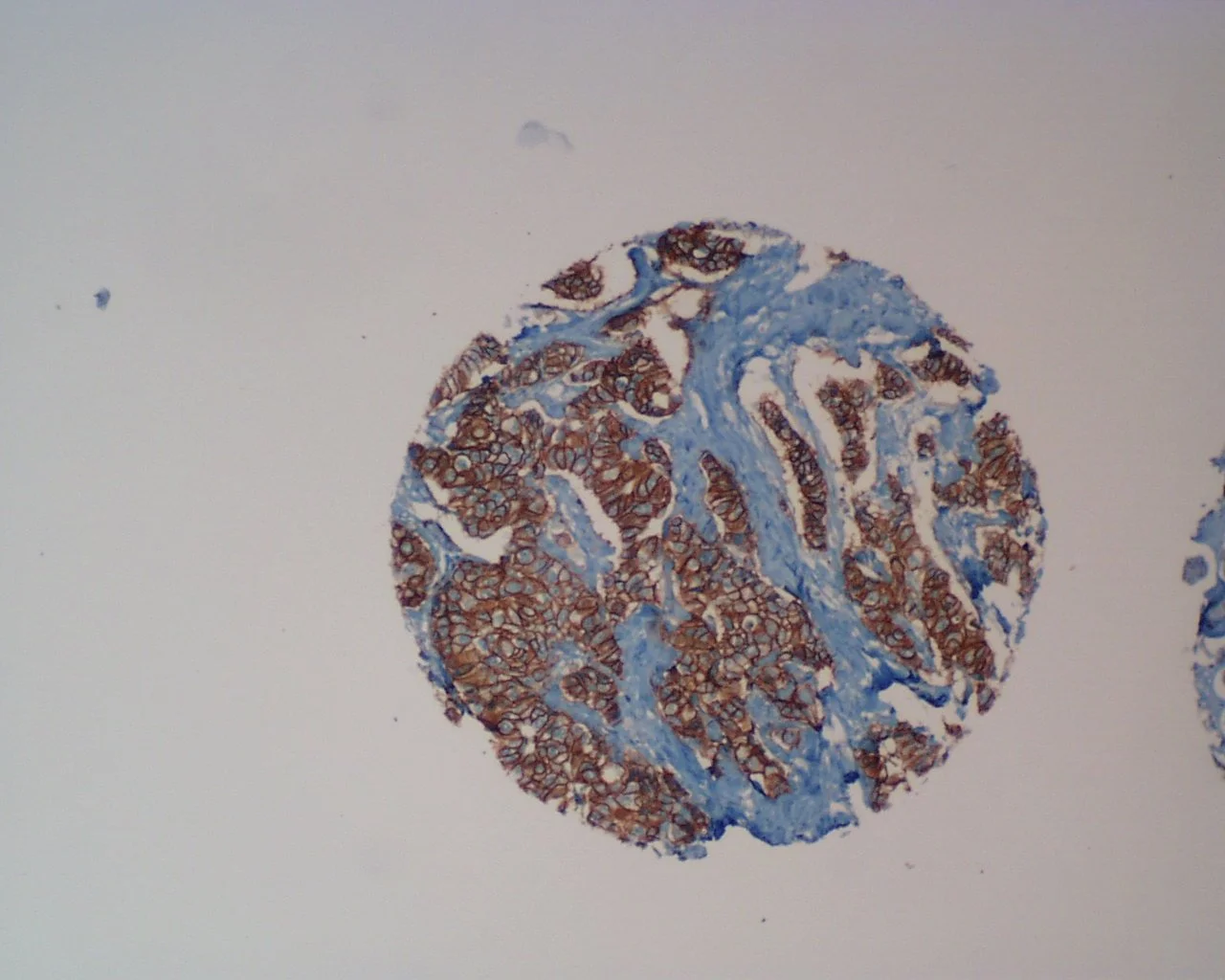
Our proprietary microarray section transfer technology assures a greater than 95% transfer efficiency. Normal tissue control sections are included on all slides.

Our Tissue MicroArrays are provided in low density format
(up to 50 tissue elements per slide)

Our Tissue MicroArrays are also provided in high density format
(up to 200 tissue elements per slide)
For each element, full pathology in clinical data is provided, some specialized arrays contain medications and treatment history. Our tissue microarrays are double spotted, representing broad surveys of specific diseases. All tissues are highly-characterized and have been carefully reviewed by board-certified Pathologists for diagnostic accuracy.

Each Tissue MicroArray is carefully designed and was crafted by highly-trained technicians. Accompanying each array set is the array locator map, and the database of clinical information (samples enclosed).
Our Tissue MicroArrays can provide your laboratory with the capability to access large datasets, for establishing expression patterns and discovering diagnostic/prognostic correlations. Your genes or proteins can be analyzed on the same set of clinical samples, enabling you to construct your own valuable expression database. Other uses can include:
Select promising gene targets
Protein profiling
Investigate disease progression and pathogenesis
Perform comprehensive molecular and proteomic profiling
WHAT WE OFFER
Click (+)to expand
-
We currently have a specialty tissue collection of both breast and prostate cancer matched with clinical data and MRI, mammography, and ultrasound data. This collection was specifically for the identification of imaging phenotypes and classifiers in correlation with tumor genotypes for characterization of breast and prostate cancer. This is a unique collection of tissue and data that may be of interest to your clients. Please inquire.
-
TMAs are created from biological materials and are such are subject to variability. For example, in tumor tissues, the amount of intervening stroma, necrosis or surrounding inflammatory reaction is highly variable. While the small size of the samples makes microarrays a powerful screening tool, the small tissue samples may not always be representative of the the entire tumor, due to the intratumor heterogeneity characteristic of most tumors. Thus, the prevalence of a molecular alteration in a specimen microarray may be underestimated. Sampling bias may not be a serious concern if the tumor areas are carefully selected for punching. This sometimes warrants collection of multiple samples from different sites of the same tumor in an attempt to obtain accurate sampling from histologically representative regions. Therefore, it is possible that some representations may be missing on particular microarray slides. For this reason, we take the time to construct our arrays to ensure a sufficient number of informative samples on every section.
-
Specimen microarrays are compiled using specimens collected in compliance with Human Subject Certification regulations, including patient consent and IRB (Institutional Review Board) Approval.